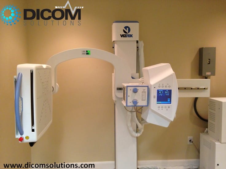
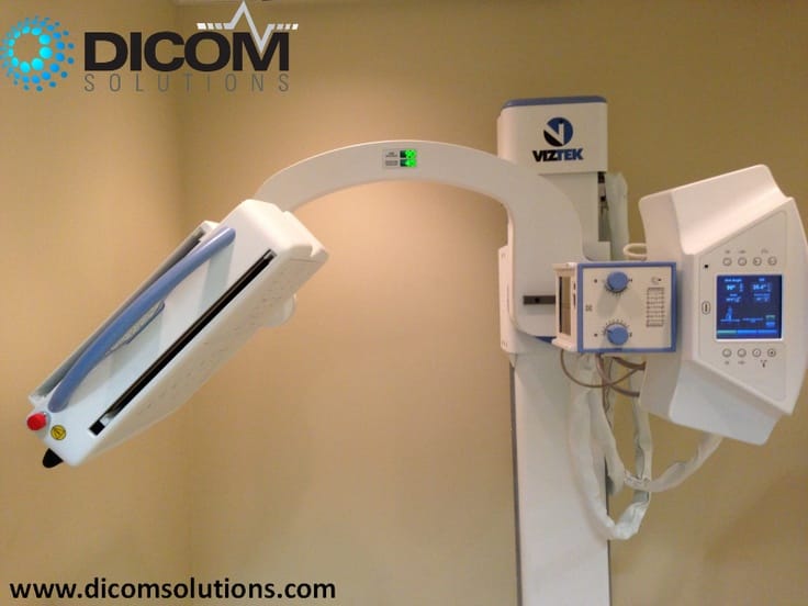
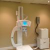
Konica 50kW U-Arm Orthopedic X-Ray
(Other kW options available) Can be combined with the DR solution best fit for your practice.
Motorized Swivel Arm with Flat Panel Cabinet
Floor-to-wall mounted support with fully motorized “U” arm for all movements. Dual Speed enables effortless and accurate settings with electromagnetic brakes for all movements.
Easy Positioning and User-Friendliness
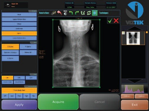
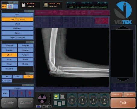
Positioning control for all movements is available from the tube/collimator head, enabling control of SID, system height, angulations for the swivel arm and detector, plus two automatic positioning buttons for chest and under table positioning. Unlimited programs are available for auto-positioning of the most common studies from the flat panel LCD 10″ color control display located on the stand. Auto Position Programming enables positioning with the push of a single button after selection of the program while with the patient.
The system (via the 10″ flat display) is able to control generator exposure parameters, enabling presetting or modification of parameters by the technician while at patient-side, before leaving to make the exposure. As the digital imaging system is fully integrated with the URS system, pre-programmed anatomical (APR) exposure techniques are synchronized with the system stand, moving the system to the corresponding position required for your study. The generator operator control console APR has been pre-programmed with the exposure parameters for various common types of study (kVp, mA, mAs and time) and mechanical position (SID, Angulations of arm and detector, height of system and collimation in case of having autocollimator) for each study, enabling auto positioning of the system with a single button.
Security Systems
Volumetric control depending on the characteristics of the room, in order to avoid possible collisions during the system positioning–anti-collision pressure sensor during the movement of the system, along the Swivel portion of the arm. Double Photocell system: One at the Start of the “U” arm area which automatically changes the speed of the system to “slow motion”, reducing the risk of harm to patient when positioned inside the arm arch. The second is located close to the end of the “U” arm area and serves to automatically “block” the system to avoid any harm or fear to the patient.
Mechanical Movements
- Variable SID distance, adjustable between 39.4 to 70.9″.
- Manual rotation of the x-ray tube +/- 180, with detents every 45 degrees
- Swivel arm range of rotation: 150, with detents on -30, 0, 90, 120 degrees.
- Swivel arm vertical movement: 50.8″.
- Minimum floor to detector center distance: 15.75″.
- Motorized Detector Angulations: +45/-45?
- Electromagnetic brakes for all movements
- Adaptation f or flat panel cabinet. Sedecal provides the mounting plate for housing for Trixel Pixium 4600 detector.
- Dimensions (H x W x D): 104.3″ x 83.1″ x 64.4″.
Manual Collimator
- Manual collimator with 6 pair of blades
- Accessory rails (cones, filters, etc)
- Field light lamp (light of the lamp 160 lux)
- Light indicator for alignment with the bucky
- Lamp timing – Retractable measuring tape
Three Phase High Frequency Generator SHF525 50kW/150kV Anatomical Program Microprocessor Controlled
- High Frequency 25KHz, 1 tube operation
- Three phase 208/240 VAC (specify prior to order)
- Output power: 640 mA @ 78kVp 500 mA @ 100kVp 400 mA @ 125 kVp 320 mA @ 150 kVp
- Automatic line compensation +/-10%
- Microprocessor controlled with auto diagnostic and error code indication for easy maintenance
- X-ray tube overload protection
- X-ray tube H.U. available indication and continuous monitoring for x-ray tube protection
- Control of x-ray tube number of exposures
- kVp Radiographic range, from 40 to 125 kVp in 1 kVp step
- mA Radiographic range from 10 to 640mA in 19 step, Renard scale 10,12.5, 16, 20, 25, 32, 40, 50, 64, 80, 100, 125, 160, 200, 250, 320, 400, 500 and 640 mA
- mAs range from 0.1 to 500 mAs in 38 steps, Renard scale
- Exposure time range from 0.001 to 10 seconds
High/Low Starter (3000/10,000 RPM) for one X-Ray tube
- Rotation control of the anode at low speed: 3.000 R.P.M., (60Hz) in function of the selected power
- Rotation control of the anode at low speed: 10.000 R.P.M., (180z) in function of the selected power
- Anode Rotation Brake control
- Time delay of the brake in function of the sequence of exposure selected
X-Ray Tube, Toshiba Model E7254FX
- Maximum Tension, 150 kVp
- Focus sizes: — Small focus 0.6 mm (0.023″) — Large focus 1.2 mm (0.047″)
- Maximum power: — Small focus 40 KW — Large focus 102 KW
- Maximum Current — Small focus 500 mA — Large focus 1000 mA
- Anode heat capacity 400,000 HU – Dissipation anode heat capacity 99,840 HU/min – Housing heat capacity 1,207,000 HU – Dissipation housing heat capacity 11,000 HU/min – Anode rotation 3,000/9,700 RPM – Anode composition Rhenium, Tungsten and face Molybdenum – Anode diameter 3.9″ – Filtration equivalent 0.028″ AL
Standard Radiographic Moving Table with brakes in two wheels; laminated structures of high compression.