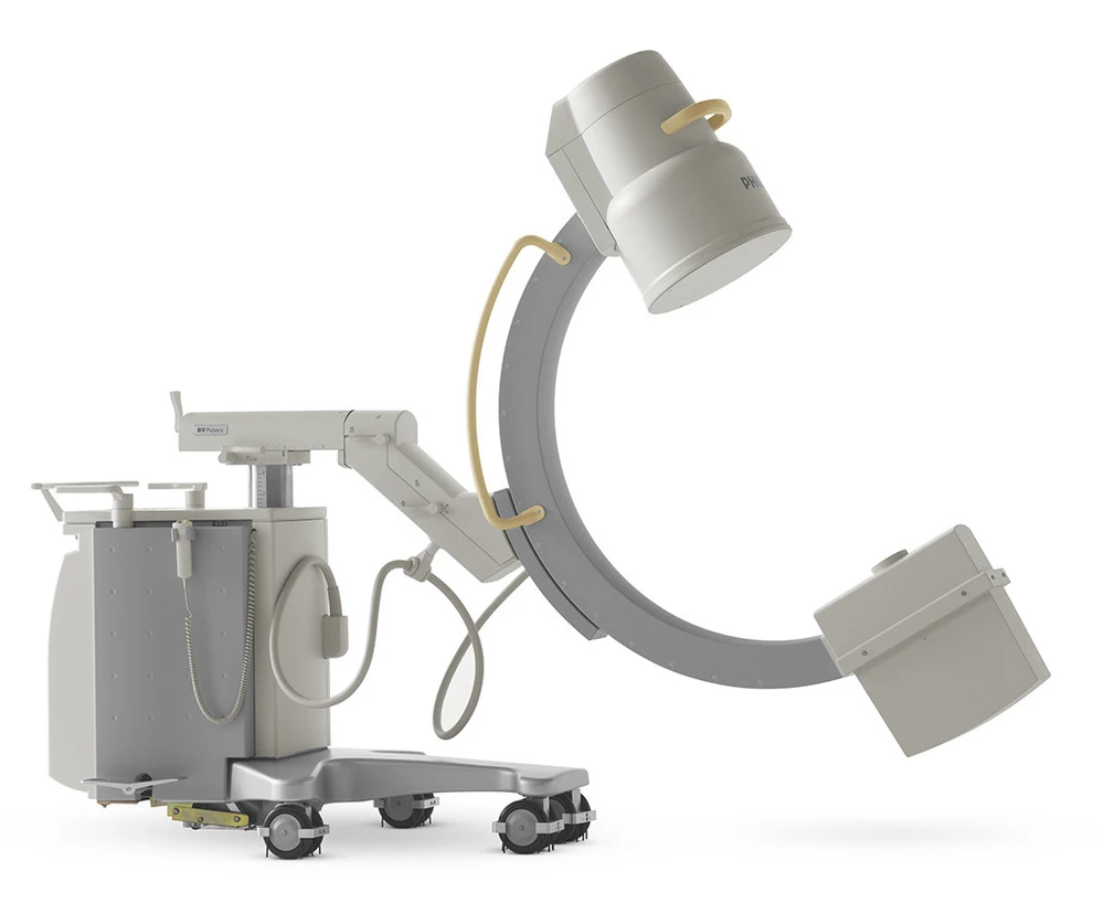
An image scanner intensifier is known as a C-arm. The C-shaped arm that connects the x-ray source and the x-ray detector is the source of the name C-arm X-ray machine.
They’re mainly employed for fluoroscopic intraoperative imaging during surgical, orthopedic, and emergency operations. The gadgets provide high-resolution X-ray images in real-time, allowing the physician to track progress and make any necessary adjustments right away.
An x-ray image intensifier (XRII) is a device that transforms x-rays into visible light at a greater intensity than conventional fluorescent panels. Such intensifiers (like fluoroscopes) are used in X-ray imaging systems to convert low-intensity x-rays into strong visible light output.
The observer may perceive the structure of the depicted item more clearly than with fluorescent displays alone because of the amplifying effect. Due to the more effective conversion of x-ray quanta to visible light, the XRII requires lower absorbed dosages.
Permanently mounted fluoroscopic devices are available in two different designs. This blog will discuss the C-arm system used where greater flexibility in the examination process is needed.
The C-arm X-ray machine is frequently employed in investigations that need a high degree of positional flexibility, such as:
- Angiography studies,
- Therapeutic studies,
- Cardiac studies,
- Orthopedic procedures.
How C-arm X-ray machines Work
A generator (X-ray source) plus an image intensifier or flat-panel detector make up a mobile fluoroscopic system, also known as a portable or mobile C-arm. The C-shaped connecting element allows for horizontal, vertical, and rotational movement around the swivel axes, allowing for X-ray imaging of the patient from practically any angle.
X-rays are emitted by the generator and penetrate the patient’s body. The image intensifier, also known as a detector, turns X-rays into a visual picture on the C-arm monitor. The physician may check anatomical features such as bones and the location of implants and equipment.
3D Imaging of a C-Arm X-Ray
C-arm computed tomography in three dimensions (3D) is a unique and revolutionary imaging technology. It generates CT-like pictures using two-dimensional (2D) X-ray projections captured with an FDP C-arm device. The C-arm system does this by doing a sweep around the subject, capturing up to several hundred 2D images.
They are used as a source of information for 3D cone-beam reconstruction. The resulting voxel data sets can be viewed as cross-sectional pictures or as 3D data sets using various volume rendering techniques. 3D C-arm imaging, initially designed for 3D high-contrast neurovascular applications, has steadily improved over time and currently gives CT-like soft-tissue picture quality. In the interventional suite, 3D C-arm imaging, when combined with 2D fluoroscopic or radiographic imaging, provides essential information for therapy planning, guiding, and outcome evaluation.
Conclusion
X-rays are crucial for the medical industry. C Arm X-ray machines are without a doubt one of the most useful medical gadgets. It assists in non-invasively diagnosing a condition or confirming a diagnosis. The physicians can review the Arm X-ray data and choose the best course of therapy or surgery.